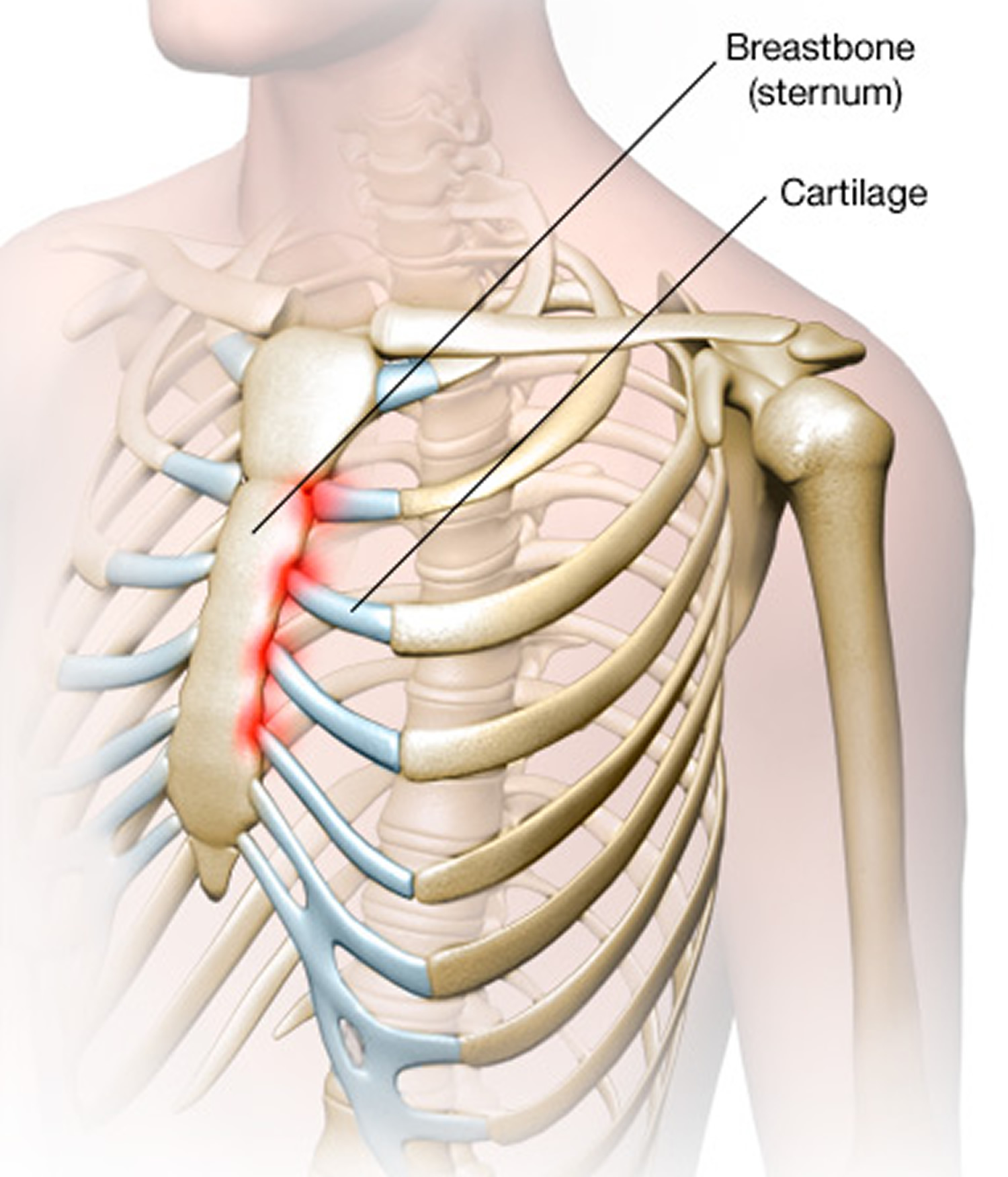
Anatomy Of Chest Wall / Figure 9 from Anatomy of the Thoracic Wall, Axilla and ... / Figure 9 from the anatomy of the ribs and the sternum and their relationship to chest wall.
byAdmin•
0
Anatomy Of Chest Wall / Figure 9 from Anatomy of the Thoracic Wall, Axilla and ... / Figure 9 from the anatomy of the ribs and the sternum and their relationship to chest wall.. Xiphoid process, costal arch, 12th and 11th ribs, vertebra t12. The chest anatomy includes the pectoralis major, pectoralis minor and the serratus anterior. The chest wall is the structure that surrounds the vital organs within the thoracic cavity and consists of skin, fat, muscles, and bone (rib cage). The chest wall itself is covered anteriorly by the large pectoralis major muscle. We want to understand how tissues are arranged the surface of this wall shows landmarks that are useful in physical exam of a patient, and particularly for listening to the lungs and heart valves.
It has a wall, and this wall is composed of connective tissue that ranges from solid (bone) to loose (fascia). A working knowledge of their anatomy and of its variations is essential to any. Anatomy of chest wall and mechanics of breathing able to describe the anatomy of the pleural cavity the pleural cavity is as if the lungs have been pushed into. Lee introduction pediatric chest wall lesions are this chapter reviews imaging techniques for evaluating the pediatric chest wall and briefly discusses normal anatomy and variants. Region in the trunk of the body that lies between the neck and…
Costochondritis - Causes, Symptoms, Locations, Duration ... from healthjade.com The chest wall is a complex system that provides rigid protection to the vital organs such as the heart, lungs, and liver; The chest anatomy includes the pectoralis major, pectoralis minor and the serratus anterior. Learn about chest wall anatomy. Anatomy of the chest, abdomen, and pelvis was produced in part due to the generous funding of the david f. Stability to arm and shoulder movement; Lee introduction pediatric chest wall lesions are this chapter reviews imaging techniques for evaluating the pediatric chest wall and briefly discusses normal anatomy and variants. Jugular notch, sternoclavicular joint, superior border of clavicle, acromion , spinous processes of c7 inferior: Anatomy, breast, axilla, chest wall, metastatic sites.
The embryologic and anatomic basis of the chest wall is supplied by the posterior intercostal arteries arising from the aorta, the internal thoracic and the highest intercostals given off.
The lobes of the lung comprise multiple bronchopulmonary segments. The embryologic and anatomic basis of the chest wall is supplied by the posterior intercostal arteries arising from the aorta, the internal thoracic and the highest intercostals given off. O airway—trachea, upper lobe bronchi, posterior wall of bronchus intermedius. How many organs could you technically live without? An understanding of chest wall kinematics might help define the loss of function after resection and the effects of various chest wall substitutes. Bones of the thoracic wall. Notice the expansile mass in the. Elastic recoil of the chest wall. Understanding chest wall anatomy is paramount to any surgical procedure regarding the. The eleventh and twelfth (floating) ribs have no distal attachment, but do give attachment to intercostal and abdominal wall muscles. Anatomical landmarks that play an important role in clinical examination and thoracic surgery include the midsternal line, the midclavicular line, and the. Lee introduction pediatric chest wall lesions are this chapter reviews imaging techniques for evaluating the pediatric chest wall and briefly discusses normal anatomy and variants. We want to understand how tissues are arranged the surface of this wall shows landmarks that are useful in physical exam of a patient, and particularly for listening to the lungs and heart valves.
Swensen fund for innovation in teaching. The chest wall, like other regional anatomy, is a remarkable fusion of form and function. Paired mammary glands, or breasts, are a distinguishing feature of mammals. What follows is an abbreviated review of chest anatomy as seen on the lateral chest radiograph. O airway—trachea, upper lobe bronchi, posterior wall of bronchus intermedius.
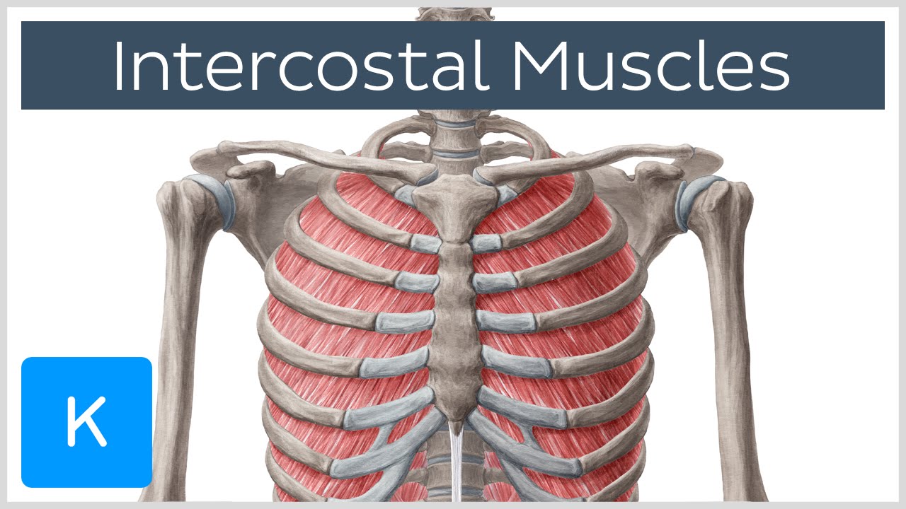
Intercostal Muscles - Function, Area & Course - Human ... from i.ytimg.com Anatomy of the chest, abdomen, and pelvis was produced in part due to the generous funding of the david f. Xiphoid process, costal arch, 12th and 11th ribs, vertebra t12. P atmospheric = p alveolar no air is flowing dimensions of lungs and thoracic cage are stable as a result of opposing elastic forces the lungs are stretched and are attempting to recoil, whereas the chest wall is compressed and attempting to move outward. Elastic recoil of the chest wall. Jugular notch, sternoclavicular joint, superior border of clavicle, acromion , spinous processes of c7 inferior: Ribs 3 through 9 are typical ribs as described earlier while ribs 1, 2, 10, 11, and 12 are atypical. The first rib is a short, flat rib that is much wider and more curved than those previously described. Region in the trunk of the body that lies between the neck and…
An understanding of chest wall kinematics might help define the loss of function after resection and the effects of various chest wall substitutes.
Anatomy, breast, axilla, chest wall, metastatic sites. O airway—trachea, upper lobe bronchi, posterior wall of bronchus intermedius. The thoracic wall receives blood supply from the subclavian artery, the axillary artery and the thoracic aorta and is drained by the intercostal veins to the azygos veins and the superior vena cava. Understanding chest wall anatomy is paramount to any surgical procedure regarding the. The chest wall, like other regional anatomy, is a remarkable fusion of form and function. Surface anatomy of anterior chest wall. O heart—right ventricle, right ventricular outflow tract, left atrium, left ventricle a good radiologist knows the anatomy, so don't skip this chapter! Stability to arm and shoulder movement; The chest wall, like other regional anatomy, is a remarkable fusion of form and function. Anatomy of chest wall and mechanics of breathing able to describe the anatomy of the pleural cavity the pleural cavity is as if the lungs have been pushed into. Region in the trunk of the body that lies between the neck and… Bones of the thoracic wall. Skandalakis je, colborn gl, weidman ta, et al.
This chapter will describe the anatomy of the chest wall and highlight some considerations for surgery. Xiphoid process, costal arch, 12th and 11th ribs, vertebra t12. Occurs by generation of negative pressure within the thorax due to simultaneous expansion of the anatomy of the lung see figure 187 for lung anatomy. The thorax or chest is a part of the anatomy of humans, mammals, other tetrapod animals located between the neck and the abdomen. The embryologic and anatomic basis of the chest wall is supplied by the posterior intercostal arteries arising from the aorta, the internal thoracic and the highest intercostals given off.
Costodiaphragmatic recess - Wikipedia from upload.wikimedia.org The chest anatomy includes the pectoralis major, pectoralis minor and the serratus anterior. Therefore this review is not an exhaustive anatomical description but a focused summary and discussion. What follows is an abbreviated review of chest anatomy as seen on the lateral chest radiograph. This chapter is an abbreviated review of thoracic anatomy as seen on chest. The chest wall, like other regional anatomy, is a remarkable fusion of form and function. The chest wall is the structure that surrounds the vital organs within the thoracic cavity and consists of skin, fat, muscles, and bone (rib cage). A complete review of the left lateral chest. Surface anatomy of anterior chest wall.
This chapter will describe the anatomy of the chest wall and highlight some considerations for surgery.
Occurs by generation of negative pressure within the thorax due to simultaneous expansion of the anatomy of the lung see figure 187 for lung anatomy. Chest wall anatomy (page 1). The chest wall is a complex system that provides rigid protection to the vital organs such as the heart, lungs, and liver; Spiral ct of thoracic inlet. Elastic recoil of the chest wall. Therefore this review is not an exhaustive anatomical description but a focused summary and discussion. Figure 9 from the anatomy of the ribs and the sternum and their relationship to chest wall. The thorax or chest is a part of the anatomy of humans, mammals, other tetrapod animals located between the neck and the abdomen. Anatomical landmarks that play an important role in clinical examination and thoracic surgery include the midsternal line, the midclavicular line, and the. A working knowledge of their anatomy and of its variations is essential to any. Region in the trunk of the body that lies between the neck and… Anatomy, breast, axilla, chest wall, metastatic sites. Anatomy of chest wall and mechanics of breathing able to describe the anatomy of the pleural cavity the pleural cavity is as if the lungs have been pushed into.
Anatomical landmarks that play an important role in clinical examination and thoracic surgery include the midsternal line, the midclavicular line, and the anatomy of chest. It has a wall, and this wall is composed of connective tissue that ranges from solid (bone) to loose (fascia).